X-PERIENCE: AN INTERACTIVE VIEWING PLATFORM DISPLAYING RADIOGRAPHIC PROFILES OF CADAVERS FOR EDUCATIONAL PURPOSES
Eithne Nic an Ríogh 1, Gary F Perry 1, James FX Jones 1, Adam Tattersall 1, Martin Heduan 1, Richard H Stern 2, Peter MacMahon 3, Nicola Giannotti 1 & Michaela Davis 1.
1 UCD School of Medicine and Medical Science, University College Dublin, Belfield, Dublin 4, Ireland
2 Radiology Department, Mount Sinai Hospital, 1468 Madison Avenue, New York
3 Radiology Department, Mater Misericordiae University Hospital, Eccles Street, Dublin 7
ABSTRACT
This project presented radiographic profiles of University College Dublin's (UCD) anatomical donors for educational purposes. An interactive viewing platform called ‘X-perience’ was developed to vividly display these images. Comprehensive donor profiles were created by adding medical histories and consultant radiologists’ reports. These image-based clinical cases enhanced anatomical learning and offered an early introduction to clinical radiography. X-perience complemented the dissection process by reinforcing the concept of the donor as the student’s first patient.
Full skeletal radiographs were obtained from 13 donors. Radiographic images were produced digitally, then labelled and stored using unique identifiers. Articulate Storyline 2 was used to build X-perience as an HTML5 interactive interface. To assess the value of X-perience, preclinical students in UCD’s undergraduate medical programme were surveyed. This cohort had prior traditional anatomical teaching and could compare anatomical learning with and without X-perience.
The workflow of profiling cadavers and constructing X-perience proved successful. X-perience is currently used by students during dissection and can be revisited via the virtual learning environment Blackboard. A survey of student opinion of the instrument (Kirkpatrick Level-1) produced favourable results. Students agreed that X-perience was relevant, easy to use and provided an integrated view of the human body. They appreciated the clinical relevance that X-perience offered. A greater understanding of the importance of radiology was acknowledged.
The introduction of similar viewing platforms in other medical schools is recommended. Imaging modalities such as CT and histology could further refine X-perience. In addition to providing educational value, X-perience strengthens the body donation programme, highlights the individuality and dignity of each donor and generates enthusiasm amongst students.
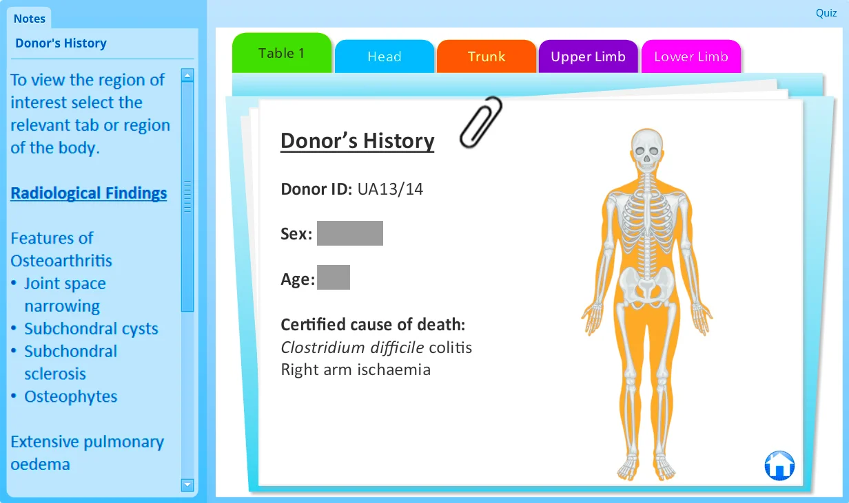
Screenshots of X-perience Interface
Article
Introduction
Traditionally, instruction in human anatomy has involved learning from both cadaveric dissection and formal lectures. The aim of this project was to introduce another dimension to learning anatomy using the medium of radiology. This integrated approach to anatomy learning was previously missing in the UCD's dissection lab. To fulfil this aim, the e-learning tool X-perience was created.
Cadaveric radiography as an anatomy teaching aid has been described in the literature as far back as 1983 by McNiesh [1] and 1985 by Pantoja [2]. More recent reports on medical student and anatomy faculty impressions of supplementing cadaver dissection with radiological images have been extremely positive [3]. Pantoja commended this approach to anatomy as an effective, easily implemented learning aid. While both McNiesh and Pantoja commented on increased student enthusiasm and interest in anatomy and radiology [1,2]. Sugand et al. state that multimodal teaching is the basis of future learning and helps combat the issue of limited dissection time [4]. While some medical schools have “abandoned dissection for user friendly multimedia”, it has been concluded that the beneficial values of orthodox dissection should not be replaced, but rather enhanced by hybrid teaching modalities [4]. A large body of research suggests that multimedia teaching is most useful in combination with dissection.
'A large body of research suggests that multimedia teaching is most useful in combination with dissection'
The Royal College of Radiologists advocates the inclusion of clinical radiology within the university curriculum [4]. Owing to the importance of radiological imaging in modern medicine, the ability of a student to interpret radiological images is vital [5]. While most medical students will not become radiologists, they must develop a fundamental knowledge of radiological imaging.6 Anatomy can be made relevant and clinically vibrant through exploration of cadaveric CT image [3]. Radiology also improves the ability to identify anatomical structures in diagnostic radiographs both in the short and long term [3]. The integration of radiographic images in the teaching of anatomy has been proven to reinforce learning and to result in better retention of both radiological and anatomical knowledge [5].
In exploring the value of examining images specific to the cadaver being dissected, Lufler et al. conclude that images need not be cadaver specific. However, cadaver specific imaging generates greater interest among students, encourages them to assume the role of a physician, and promotes respect for donors [7].
X-perience presents donor specific radiographic profiles of UCD cadavers for educational purposes. This interactive viewing platform displays radiographic images, giving UCD anatomy students the opportunity to explore interesting findings and common pathologies relating to specific donors.
Please note that identifying details have been redacted for purposes of publication.
Methods
Commencing in May 2015 the diagnostic imaging department of UCD obtained full skeletal radiographs from 13 donors (10 Female and 3 Male) aged 58-101 years. Each donor was transferred from the anatomy laboratory to the imaging room where images were captured. Radiographic images consisted of antero-posterier (AP) and lateral skull, cervical spine, chest, upper and lower arm, hand, abdomen, pelvis, upper and lower leg. This provided a complete skeletal survey of each donor. The images were produced digitally using Digital and Computed Radiography and stored securely as a DICOM dataset using unique identifiers for each donor. Exposure factors and total radiation dose were recorded.
Using Adobe Photoshop (Adobe, United States) the identity was removed from each image and the images were systemically labelled. Collaborating with consultant radiologists in Mount Sinai Hospital, New York and in the Mater Misercordiae Hospital, Dublin the images were systematically labelled with both anatomical and pathological findings. This made the images clinically relevant for students as well as a valuable study tool for anatomy. Khalil et al. stress the importance of clear labelling to empower rather than overwhelm the student [6]. Clinical histories were researched from donor files and presented alongside the images to provide a comprehensive profile on each donor.
The e-learning authoring software Articulate Storyline 2 (Articulate Global Inc., United States) was used to construct X-perience. This software allowed for the creation of a web enabled HTML5 interactive interface that students were able to use on the touch screens in the dissection lab. Students were initially presented with a welcome screen on which they were reminded of the sensitive nature of the material they were viewing. Following this, a simple map of the dissection room was displayed from which students could select the donor whose profile they wished to view. An interactive folder displayed the donor history as well as an interactive body outline from which the relevant body region could be selected. The images were grouped according to four body regions: head, trunk, upper limb and lower limb.
The body region of interest could be chosen by clicking on the body outline or by clicking on the tabs. Factual information was presented alongside the images of interest, briefly describing topics such as intramembranous and endochondral ossification, fracture classification and treatment options. On viewing a specific image, students were initially presented with an unlabelled image, which allowed feature discovery by the student. Labels pointing out relevant anatomical structures in green and pathological findings in pink could be turned on as desired. The radiological findings relevant to the specific image could be seen alongside the image in a side bar.
“This drag and drop quiz allowed students to test their anatomy knowledge”
Another valuable feature of X-perience was the interactive quiz. This drag and drop quiz allowed students to test their anatomy knowledge. When a correct label was dragged from the bottom of the screen to the relevant region, the label slotted automatically into place. Incorrect answers returned automatically to the bottom of the screen. This easy to use quiz provided instant feedback to students and was a fast and effective method of testing anatomy learning.
To assess the value of X-perience preclinical students in UCD’s undergraduate medical programme, specifically Stage 3 Locomotor Biology students (n = 50) were surveyed. This cohort had prior traditional anatomical teaching in Stage 2, and thus could effectively compare anatomical learning with and without X-perience. Anatomy laboratory demonstrators were also consulted for feedback on X-perience. Responses were recorded using a 5 point Likert scale of strongly agree, agree, neutral, disagree and strongly disagree.
The following statements were presented on the survey:
- I found X-perience easy to use.
- X-perience is helpful for learning anatomy.
- X-perience helps me understand why anatomy is the foundation of much of my coursework.
- X-perience gives me a better appreciation of my anatomical donor.
- I now have a better understanding of the clinical application of radiology.
- X-perience is relevant to medical education.
- I will access X-perience via Blackboard to revise the material.
- I can identify anatomical structures more accurately after using X-perience.
- X-perience helps bring material from numerous modules together, creating an overall picture of the human body.
An open-ended response and comment section was also included where students were encouraged to give feedback.

Results
The workflow of profiling cadavers and constructing X-perience proved successful.
X-perience is currently used by medical students on touch screens during practical classes and can be revisited via the virtual learning environment Blackboard. The results of the student acceptability survey, Kirkpatrick Level 1, showed extremely positive student feedback. 84% of students agreed that X-perience was a relevant, helpful and easy to use learning tool. 84% of students found that X-perience provided them with a more integrated, complete view of the human body and brought material from numerous modules together. Students (88%) appreciated the clinical relevance that X-perience brought to their learning. A greater understanding of the importance of radiology was acknowledged by 92% of students. 92% of students felt that by using X-perience they gained better appreciation of the anatomical donors and their medical histories. Students’ reported comments were: “Absolutely brilliant”, “Very cool”, “Extremely interesting and useful”, “Very impressive software”.
'Very Cool'
'Absolutely Brilliant'
'Extremely Interesting'
Conclusions
While radiological profiles of donors have been carried out as far back as 1983, many medical schools have yet to adopt this learning tool. Following the successful integration of radiological profiles in UCD anatomy teaching, the introduction of similar viewing platforms at other medical schools can be recommended. The unique benefit of X-perience is the interactive, student-friendly display of the clinical profiles. The method of display encourages students to delve more deeply into their anatomy learning and explore the individuality of their donor. Advanced imaging modalities such as CT, as well as histology, could further refine X-perience and it is hoped that this will be carried out in future years. In addition to its educational value, X-perience strengthens the body donation programme, highlights the individuality and dignity of each donor and generates enthusiasm amongst anatomy students.
ACKNOWLEDGEMENTS
We would like to acknowledge the generosity of all the anatomical donors to UCD for providing us with the opportunity to learn in such a unique and privileged way.
Exemption from full ethical review was granted by the Human Research Ethics Committee – Sciences in University College Dublin.
References
- McNiesh LM, Madewell JE, Allman RM. Cadaver radiography in the teaching of gross anatomy. Radiology. 1983 Jul;148(1):73–4.
- Pantoja E, Nagy F, Zambernard J. Clinical radiographs of the cadaver as a teaching aid in anatomy. Radiology. 1985 Apr;155(1):28.
- Bohl M, Francois W, Gest T. Self-guided clinical cases for medical students based on postmortem CT scans of cadavers. Clin Anat N Y N. 2011 Jul;24(5):655–63.
- Sugand K, Abrahams P, Khurana A. The anatomy of anatomy: a review for its modernization. Anat Sci Educ. 2010 Apr;3(2):83–93.
- Rengier F, Doll S, von Tengg-Kobligk H, Kirsch J, Kauczor H-U, Giesel FL. Integrated teaching of anatomy and radiology using three-dimensional image post-processing. Eur Radiol. 2009 Dec;19(12):2870–7.
- Khalil MK, Payer AF, Johnson TE. Effectiveness of using cross-sections in the recognition of anatomical structures in radiological images. Anat Rec B New Anat. 2005 Mar;283(1):9–13.
- Lufler RS, Zumwalt AC, Romney CA, Hoagland TM. Incorporating radiology into medical gross anatomy: does the use of cadaver CT scans improve students’ academic performance in anatomy? Anat Sci Educ. 2010 Apr;3(2):56–63.